DEVICE FOR THE TREATMENT OF ANISEICONIA WITH MAGNETISED GLASSES IN BRIDGE
|
Descripción |

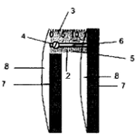
Diagram of the device assembly: metal bar (2) millimetre ruler (3) screws (4) cutting of the cover (5) slit (6) eyepieces (7) lenses (8)
It is a device that allows the treatment of aniseiconia or retinal image difference through magnetized glasses in its bridge, favoring the magnification of the retinal image of smaller size until equality with that of the other eye. The position is measured with differences in mm in which the aniseiconia disappears.
|
¿Cómo funciona? |

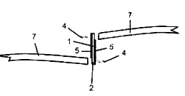
Side face of the device before mounting: magnetic bridge glasses (1)
Aniseiconia (a condition of binocular vision in which there is a relative difference in the size and/or shape of the perception obtained from the retinal image when transmitted to the visual cortex) can be compensated for by variations in the mount and its adaptation. To compensate for the aniseiconia, the corresponding eyepiece of the magnet glasses, already marketed, is moved and the device is atomized, the cover is cut at both ends glued to the eyepieces, the bar is also cut at the ends so that it does not protrude, polishing them properly.
|
Ventajas |
The greatest advantage is the similar value of the accommodation amplitude to that obtained with the negative lens procedure. This new device eliminates test errors such as overestimating or underestimating the accommodation amplitude. In addition, the increase of the optotype is avoided. The major advantage is to provide equality in retinal images avoiding fusion problems and all the symptomatology involved (headache, tearing, asthenopia, etc..) with a commercial model of glasses with split bridge. This new device allows the treatment of aniseiconia without the use of iseiconic lenses impossible to find on the market due to the difficulty of manufacturing on a normal scale given the low incidence of this problem in the population.
|
Donde se ha desarrollado |
The design, protected by national patent with prior examination since 2013, and its prototype, have been developed in the Faculty of Optics and Optometry.
|
Contact |
|
© Office for the Transfer of Research Results – UCM |
|
PDF Downloads |
|
Classification |
|
Responsible Researcher |
Ricardo Bernárdez Vilaboa: rbvoptom@ucm.es
Department: Optometry and vision
Faculty: Optics and Optometry


