TRIAL FRAME FOR MEASURING ANISEICONIA
|
Description |
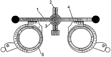
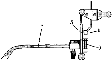
Front outline of the frame: 1. Frame. 2. Cut on the bridge of the saddle. 3. Rule. 4. Naso pupillary distance scale. 5. Eyepiece
It is an apparatus that evaluates the perception of images in the retinas of both eyes and allows correcting it in case of anomaly or defect of focus.
The main feature of the device is that it has a frame that allows to separate one half of the device from the other. Thus, each separate eyepiece has the possibility of inserting up to 3 lenses of different calibers for the compensation of refractive error. Each lens is placed in different positions to ensure that the distance between the lenses and the eye (distance to the vertex) is different. With this change in distance, the aim is to equalize the retinal image in both eyes, by means of a rule that measures the separation of both oculars in mm.
|
How does it work |
There are several methods to evaluate aniseiconía (ocular alteration caused by a difference in the size and / or shape of the perceived images). This device modifies a spectacle frame to change the size of the retinal image.
To perform the measurement that allows the best equality of the retinal images, the eyepiece is moved away with the lens of greater positive power or of less negative power until the patient indicates that he perceives the two images of the same size. At that exact point we can read in the rule the separation in mm that we have between the two eyepieces.
This new equipment allows to modify the distance to the vertex, in addition to enabling the rest of the base curve and central thickness adjustments of the lenses.
|
Advantages |
• Simplification of the distance to the vertex by providing equality in the images.
• It avoids problems of fusion and the symptomatology that it entails (headache, tearing, asthenopia, etc.)
|
Where has it been developed? |
The design, protected by national patent with previous examination since 2013 has been developed in the Faculty of Optics and Optometry.
|
Contact |
|
© Office for the Transfer of Research Results – UCM |
|
PDF Downloads |
|
Classification |
|
Responsible Researcher |
Ricardo Bernárdez Vilaboa: rbvoptom@ucm.es
Department: Optometry and vision
Faculty: Optics and Optometry


