BONE REGENERATION WITH CALCIUM CEMENTS AND CERAMICS (BRUSHITA, MONETITA, Β-TCP). IN VITRO AND IN VIVO TRIALS USING A DEVICE MODEL IN RABBIT'S FOOTPRINT
|
Description |

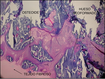
(1). 10% Fe-β-TCP ceramics at 8 weeks with neoformed bony trabeculae and a large osteoblastic forehead.
In many cases in dentistry patients are found that have insufficient bone availability, either for the placement of osseointegrated implants or dental prostheses. Also in the case of some periodontal diseases, tissue loss occurs and healthy bone formation is required. To solve these problems, regenerative strategies are used to reconstruct the atrophic maxilla. There is currently a wide range of calcium phosphates for use in bone regeneration procedures. Calcium phosphates are of particular interest because they resemble the mineral phase of human bone and are susceptible to bone remodeling and reabsorption, with the most used being hydroxyapatite, beta-tricalcium phosphate (β-TCP) and monetite. These materials have demonstrated a great regenerative capacity especially in retentive cavities, such as the dental alveoli or inside the maxillary sinus. Calcium phosphate cements are biocompatible synthetic materials, which promote bone regeneration, decompose under physiological conditions and are reabsorbed by the body. The calcium cement of brushite and monetite have been shown to be well accepted by the body and favor bone regeneration. The chemical composition of these materials is suitable to create porous structures that allow cell proliferation and formation of blood vessels during the process of bone regeneration. In our laboratory we have developed the methodology to use a combination of gels and polysaccharides to control the properties of brushite and monetite cements. In in vivo studies with rabbit animal models we have investigated the bone regeneration of our cements and the histological observation of the grafted samples of cement granules reveals a good resorption of the cement and a consequent filling of the space with new bone formed. Recently, synthetic granular materials of brushite and monetite have been evaluated in both animal and human models and appear as an alternative to the use of autogenous bone since very good results are obtained both in obtaining regenerated bone and in resorptive capacity in retentive cavities. We are also conducting studies with membranes based on hybrid polymer materials interpenetrated with calcium phosphate particulates for guided bone regeneration.
|
How does it work |
- Calcium phosphate microparticles. Microparticles of calcium phosphate cements are synthesized by mixing a solid phase with another liquid which may have growth factors. The materials are presented as granules with a particle size of about 200 microns which prevents their diffusion into the bloodstream and the formation of thrombi. Optimized porous matrices can be obtained to promote bone regeneration and angiogenesis (Figure 1).
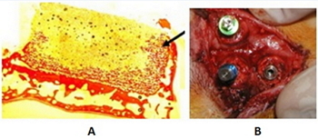
- Bolted blocks of monetite. We developed solid 3D matrices of monetite with sufficient rigidity and a design that allows its fixation to the bone by means of screws of osteosynthesis without being fractured, with a suitable porosity to allow the cellular growth, and with capacity of biorreabsorción to facilitate the processes of bone remodeling (See Figure 2).
- Calcium phosphate ceramics replaced by strontium, magnesium, iron and silicon ions. Ionic substitution is a tool to modify the physico-chemical and biological properties of osteoconductive ceramics of β-TCP. The incorporation of strontium, magnesium, iron and silicon ions is useful for obtaining a bone substitute with better properties without compromising the biocompatibility of the starting material. For example, strontium ions improve the hardness of the material and iron ions provide the material with magnetic properties without altering its biological response (Figure 3).
- In vitro trials of cellular cytotoxicity in biomaterials. For the study of cytotoxicity and cell proliferation in biomaterials, cells from the human cell line are cultured in a culture medium. The viability of the cells is evaluated by cell proliferation reagent WST-1. Cell proliferation is calculated by electronic cell count.
- Formation of hybrid polymer membranes and particles of calcium phosphate. Polymer membrane mineralization (poly (ethylene oxide), agarose, carrageenan, etc.) will be used in the process of diffusion of reagents followed by chemical reaction and formation of precipitates. Reagent diffusion can be performed either by concentration gradient or by using a potential gradient.
- Antimicrobial activity of ceramics and cements. The antimicrobial activity in vitro allows to determine the sensitivity of the microorganism to the antimicrobial agent and its bacteriostatic potency.
- In vivo assays of biomaterials: Histological and histomorphometric studies.

(2) A) Microscopic image of calcium phosphate cement consisting of brushite microcrystals. B) Mesenchymal cells growing within a pore of a porous matrix of brushite cement. C) Bone regeneration promoted by a brushite cement granule (dark brown) surrounded by a non-mineralized (purple) bone matrix and mineralized immature bone (light purple) which indicates that bone formation follows the reabsorption of the brushite granule.
|
Advantages |
The profitability of obtaining biocompatible materials that promote the bone regeneration allows:
The reduction of production costs:
- Chemical synthesis of bone regeneration products.
- Use of biocompatible and biodegradable products
- The design of solid arrays.
- Assurance of its bioavailability.
- Preparation of materials à la carte.
In relation to the distribution:
- Easy handling and application.
- Easy storage.

(3) A) Histological image of a 3D block of 4mm high monetite, screwed onto a rabbit's cap, 4 weeks after placement. Bone neoformation is observed along the whole block, reaching even the uppermost region on the right side (black arrow). B) Image of a rabbit's vault at the time of placement of endosteal implants inside the monetite blocks, showing excellent stability.
|
Where has it been developed |
The Bioactive Nanostructured Materials Research Group (MATNABIO) of the Department of Physics-Pharmaceutical Chemistry of Faculty of Pharmacy has developed methods of synthesis of materials based on calcium phosphates and also possesses techniques of characterization of these systems. Our team has demonstrated that it is able to develop new application biomaterials in bone regeneration procedures as reflected in the publication of articles in high-impact journals and in the development of patents.
|
And also |
The research group has experience in collaborations with private companies in the polymer industry, biomaterials and dental materials. And it has the following related patents:
-Brushite as an enzyme immobilization system and its use in biocompatible amperometric biosensors.
-Stable biocompatible stabilizers for immobilization of enzymes and their use in amperometric biosensors.
-Polymer microgel particles with immobilized glucose oxidase and their use as a glucose biosensor.
-Synthetic blocks for use in dental implants.
-Cement of calcium phosphate - calcium silicate for biomedical applications.
|
Contact |
|
© Office for the Transfer of Research Results – UCM |
|
PDF Downloads |
|
Classification |
|
Responsible Researchers |
Enrique López Cabarcos: cabarcos@ucm.es
Carmen Rueda Rodríguez: crueda@farm.ucm.es
Jesús Torres García-Denche: jesus.torres@ucm.es
Department: Pharmaceutical Physical-Chemistry
Faculty: Pharmacy


