Modificacion de superficies de óxidos
The search for functionalities and improvements of the physico-chemical properties of many materials often requires a previous treatment involving surface modifications. Low energy ion bombardment is a common tool for this purpose, since the incoming ions have a sufficiently low energy to affect only the topmost layers of the material. The energy is, however, high enough to promote drastic changes in the topography and chemical composition. And some relevant physical and chemical properties (catalysis, adhesion, wettability...) depend mostly on the nature of these topmost layers of the material.
Oxide surfaces are very interesting targets for ion-induced modifications, as their tolerance to non-stoichiometry allows the exploration of a wide range of induced morphologies and properties. Within this research line we have studied
a) the transformation of the surface of a TiO2(110) surface into a single-crystalline thin film (10 nm) of TiO(001) (reference [1] below).
b) the fabrication of a single crystalline Fe3O4(111)/Fe2O3(0001) bilayer (references [2] and [3] below).
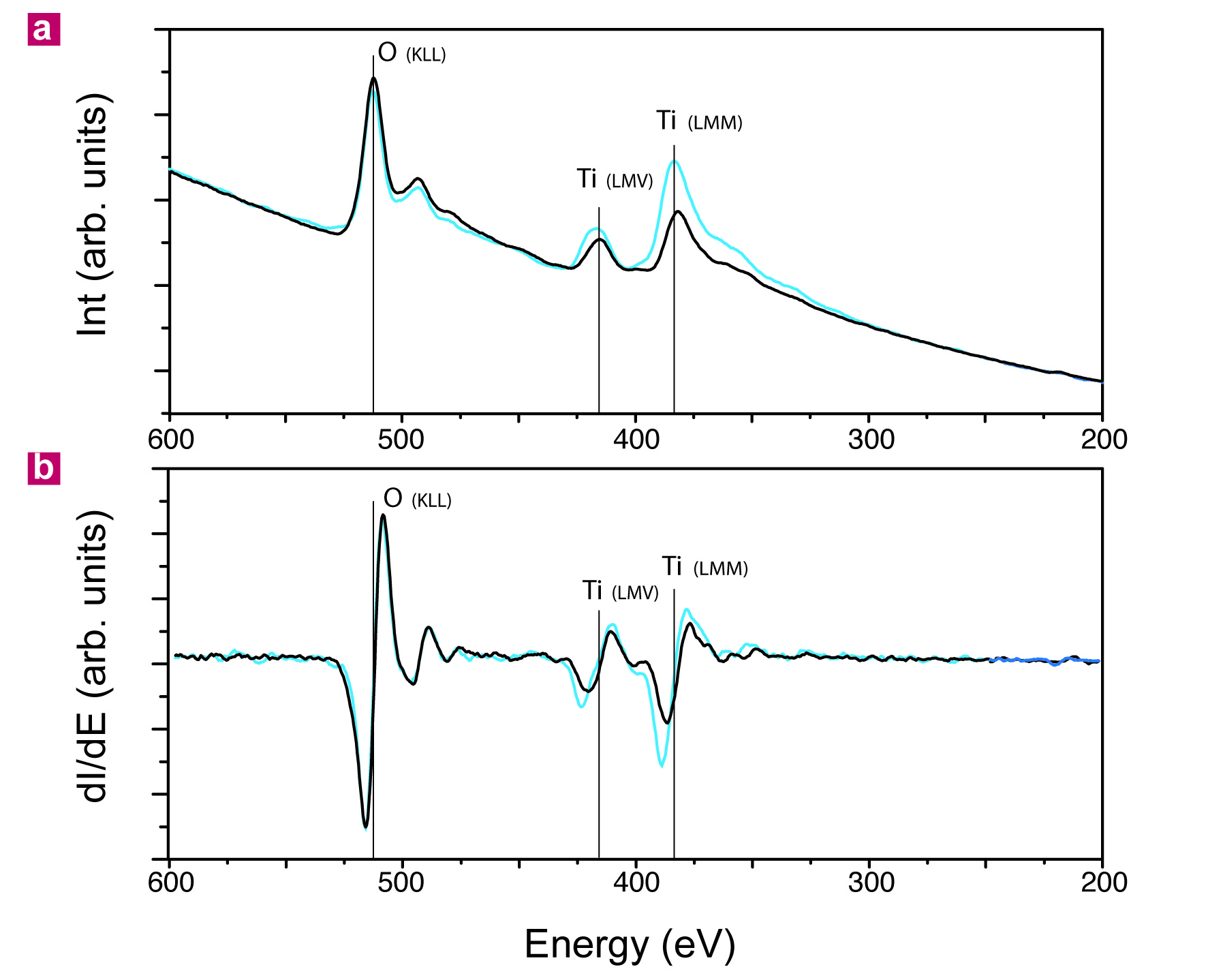
The Auger spectra below shows, for instance, the chemical modification (reduction) of the TiO2(110) surface after ion bombardment (blue curve, as compared to the pristine surface in black).
The maps in reciprocal space below show the coexistence of two crystal lattices after ion bomabrdment: the black lattice in the HK maps on the left correspond to the TiO2(110) surface, while the blue lattice belongs to the TiO(001) lattice, a product of ion bombardment. L-scans (below on the right) also show the out-of-plane periodicity.

The main conclusion of this work is that low energy ion bombardment can be used to produce a single-crystalline thin film of a suboxide on top of the corresponding oxide.

XMCD (a) and DF-LEEM (b) images of the same region of the magnetite surface. This magnetite phase has been produced by low energy ion bombardment of a hematite epitaxial thin film [3]. The red ellipse on the bottom left is the same exact location on the surface. A smooth dichroic contrast is obtained (with darker and brighter regions). c) XMCD spectra taken at different helicities (σ+ and σ−) integrated on a specific area of the magnetite surface. The black curve (the dichroic spectrum) is a subtraction of both helicities. The insert is an XMCD image, and the small yellow area (with a dark dichroic contrast) is the exact location where both spectra have been integrated. The XMCD images from (a) and (c) have been taken at different locations of the surface.
To our best knowledge, these are the first examples of how low energy ion bombardment can be used to transform an oxide surface into a single-crystalline thin film of the corresponding suboxide. Our work clarifies the role of ion bombardment on the atomic structure and establishes a new route to generate, for certain cases, heterostructures and interfaces between an oxide and its corresponding suboxide, in a relatively simple and controlled way. Low energy ion bombardment shows in this case a new technologically relevant capability.
These results have been published in published in Nature Communications, Applied Physics Letters and Ultramicroscopy:
[1] “Formation of titanium monoxide (001) single-crystalline thin film induced by ion bombardment of titanium dioxide (110)”
B.M. Pabón, J.I. Beltran, G. Sanchez-Santolino, I. Palacio, J. Lopez-Sanchez, J. Rubio-Zuazo, J.M. Rojo, P. Ferrer, A. Mascaraque, M.C. Muñoz, M. Varela, G.R. Castro and O Rodríguez de la Fuente
Nature Commun. 6 6147, (2015)
DOI: 10.1038/ncomms7147
[2] "Formation of a magnetite/hematite epitaxial bilayer generated with low energy ion bombardment"
S. Ruiz-Gómez, A. Serrano, I. Carabias, M. A. García, A. Hernando, A. Mascaraque, L. Pérez, M. A. González Barrio, and O. Rodríguez de la Fuente
Appl. Phys. Lett. 110, 093103 (2017)
DOI: 10.1063/1.4977491
[3] "Spectromicroscopic study of the transformation with low energy ions of a hematite thin film into a magnetite/hematite epitaxial bilayer"
Mauricio J. Prieto, Lucas de Souza Caldas, Liviu C. Tănase, Thomas Schmidt, Óscar Rodríguez de la Fuente
Ultramicroscopy 255, 113855 (2024)
https://doi.org/10.1016/j.ultramic.2023.113855